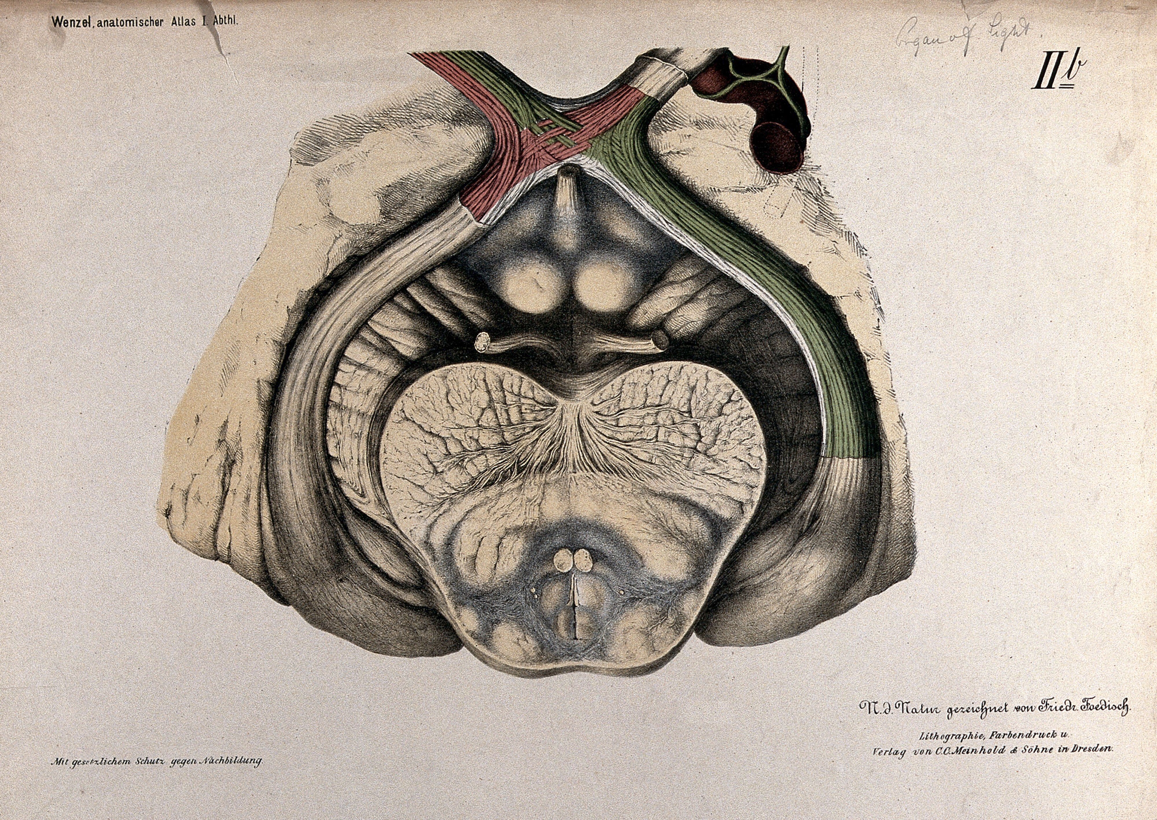
Anatomical section of the brain showing the optic chiasma. Colour lithograph by F. Foedisch, 1875.
A colour lithograph of a brain seen from below, showing the cerebellum's folded surface in the lower center and red and green nerve fibres curving across the top, including the optic chiasm. Dark shading and pale tissue give the plate a scientific, atlas-style look.
- Artist
- Foedisch, Friedrich, active 1875-1877.
- Date
- [1875]
- Medium
- Lithographs
- Source
- Wellcome Collection
- Size
- 4000 × 2835 px
Rights checked 2026-11-05 · tier A · as stated by the source: Public Domain Mark (Wellcome Collection)
Credit (appreciated, not required for public-domain works): Wellcome Collection
Español: Anatomía de la base del cerebro con quiasma óptico, litografía de 1875
Litografía en color de un cerebro visto desde abajo, con la superficie plegada del cerebelo en el centro inferior y fibras nerviosas rojas y verdes que cruzan la parte superior, incluido el quiasma óptico. Las sombras oscuras y los tejidos pálidos dan a la lámina un aspecto científico de atlas.
Português: Anatomia da base do cérebro com quiasma óptico, litografia colorida de 1875
Litografia colorida de um cérebro visto por baixo, com a superfície dobrada do cerebelo no centro inferior e fibras nervosas vermelhas e verdes que cruzam a parte superior, incluindo o quiasma óptico. As sombras escuras e os tecidos claros dão à prancha um aspecto científico, de atlas.